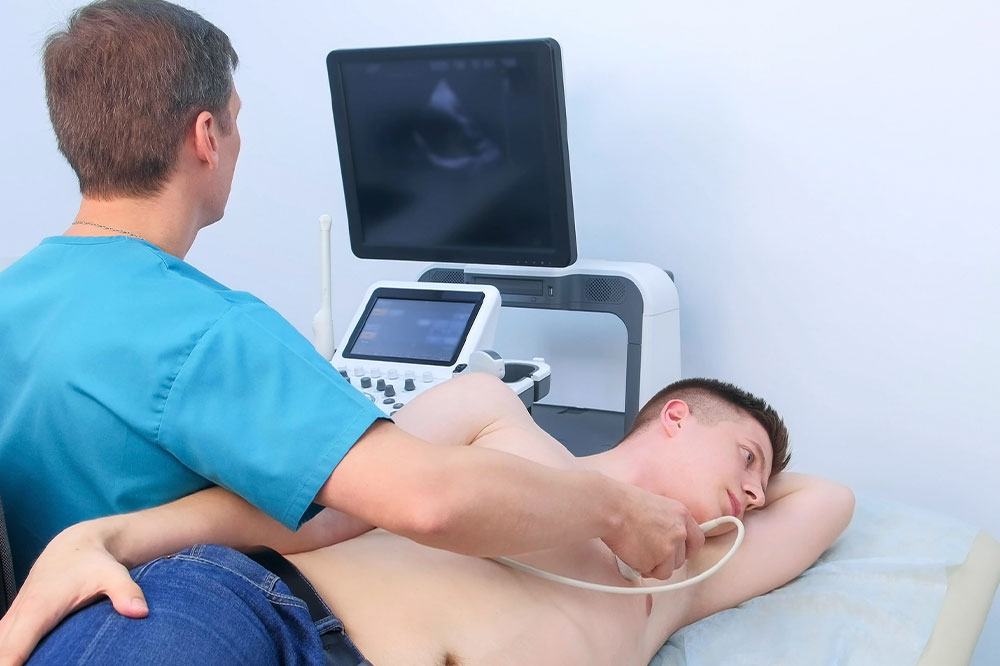
Echocardiography – Benefits, types, and steps involved
Echocardiography is a diagnostic test that uses sound waves to create live pictures of the heart. These pictures, called echocardiograms, provide important information about the organ’s chambers, valves, blood flow, and overall function. They are more detailed than ordinary X-ray images. Echocardiography helps doctors evaluate the condition of a person’s heart muscle, especially after a heart attack. It can also detect heart problems in unborn babies, making it incredibly helpful.
What are the benefits of echocardiography?
Echocardiography helps doctors assess heart health. It can screen people at risk of heart disease, monitor the development of a baby’s heart, track treatment progress, and assist in surgery. It can also help identify heart problems like:
- Heart failure
- Heart valve issues
- A heart that is larger than usual or has thickened ventricles (lower chambers)
- Weakened heart muscles
- Heart defects present since birth
- Blood clots or tumors in the heart
- Artery blockages
What are the types of echocardiography?
There are different ways of performing the test. Here are a few common examples:
- Transthoracic echocardiography (TTE)
A device is placed on the patient’s chest to capture heart images. It provides detailed information about the organ’s structure, chamber size, valve condition, and how well it pumps blood. Simple and painless, TTE is widely used in practice. - Transesophageal echocardiography (TEE)
A probe is inserted into one’s esophagus to visualize the heart better. It is useful when TTE images are unclear or when a more comprehensive examination is required. TEE tests detect blood clots, infections, or heart abnormalities during surgeries. - Stress echocardiography
This test captures heart images before and after exercise or treatment. It helps diagnose coronary artery disease by showing if parts of the heart are not receiving enough blood during exercise. It also evaluates exercise capacity, detects irregular heartbeats, and checks the effectiveness of treatments. - Three-dimensional echocardiography (3DE)
It creates 3D images of the heart using special tools and advanced imaging techniques. The 3D images help doctors analyze the heart’s structures more accurately. 3DE helps assess complex heart problems, including issues with the organ’s structure, valves, and congenital heart disorders. - Fetal echocardiography
This special test allows doctors to monitor the baby’s heart while still in the mother’s womb. A transducer placed on the mother’s belly captures images of the baby’s heart. It helps identify problems before or after birth, enabling doctors to provide appropriate counseling and care.
While these procedures are usually harmless, some experience minor discomfort or have allergic reactions to the contrast solutions. Hence, one should talk to a doctor about allergies or sensitivities before the test.
A look at the echocardiography procedure
Preparation for a TTE is simple. Patients are asked to avoid eating or drinking anything for a few hours before the test. Here’s what a TTE procedure looks like:
- Patients undress up to the waist and lie on their backs on a testing table.
- Electrodes are placed on the chest to track heart rate.
- A tiny amount of gel is applied to the chest.
- A transducer is moved softly across the skin (it may exert minor pressure).
- The patient might be asked to breathe a certain way or roll onto their left side.
Those undergoing a TEE are administered a solution before the probe is inserted. A numbing fluid may also be sprayed on the back of the throat.


